B-Rpe65 KO mice

    C57BL/6JNifdc-Rpe65tm1Bcgen/Bcgen • 113758

    B-Rpe65 KO mice

    Product nameB-Rpe65 KO mice
    Catalog number113758
    Strain nameC57BL/6JNifdc-Rpe65tm1Bcgen/Bcgen
    Strain backgroundC57BL/6JNifdc
    NCBI gene ID (Mouse)
    AliasesLCA2; RP20; rd12; 65kDa; Mord1; A930029L06Rik

    在此页面上

    • Description
    • Targeting strategy
    • Phenotypic analysis

    海报

    查看全部

      发表文章

        Description

        Background:

        • The human RPE65 gene is located at 1p31 and encodes the retinal pigment epithelium-specific 65 kDa protein (RPE65). As a key enzyme of the carotenoid oxygenase/lyase superfamily, it plays an essential role in retinal vitamin A metabolism.
        • RPE65 is a membrane-bound protein featuring an N-terminal signal peptide and a C-terminal esterase domain. Its three-dimensional structure comprises multiple transmembrane helices and extracellular loops. RPE65 primarily catalyzes the hydrolysis and isomerization of all-trans retinyl esters in the visual cycle, facilitating the regeneration of 11-cis retinol (the chromophore of rod and cone opsins).

        Targeting strategy:

        • The exons 3-15 of mouse Rpe65 gene were knocked out in B-Rpe65 KO mice.

        Verification: 

        • Mouse RPE65 protein was only detected in the eye of C57BL/6JNifdc mice, but not in homozygous B-Rpe65 KO mice.  
        • Mouse Rpe65 mRNA was only detectable in wild-type mice, but not in homozygous B-Rpe65 KO mice.

        Application:

        • This product can be utilized to assess the pharmacodynamic effects of therapies targeting autosomal recessive retinitis pigmentosa (RP) and Leber congenital amaurosis (LCA).
        Targeting strategy

        Gene targeting strategy for B-Rpe65 KO mice. The exons 3-15 of mouse Rpe65 gene were knocked out in B-Rpe65 KO mice. As a result, mouse RPE65 protein will not be expressed anymore.

        Protein expression analysis

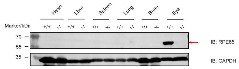
        Western blot analysis of RPE65 protein expression in homozygous B-Rpe65 KO mice. Various tissue lysates were collected from wild-type C57BL/6JNifdc mice (+/+) and homozygous B-Rpe65 KO mice (-/-), and then analyzed by western blot with anti-mouse RPE65 antibody (Abcam, ab231782). 40 μg total proteins were loaded for western blotting analysis. Mouse RPE65 was only detected in the eye of C57BL/6JNifdc mice.

        mRNA expression analysis

        Strain specific analysis of mRpe65 mRNA expression in wild-type C57BL/6JNifdc mice and B-Rpe65 KO mice by RT-PCR. Eye RNA were isolated from wild-type C57BL/6JNifdc mice (+/+) and homozygous B-Rpe65 KO mice (-/-), then cDNA libraries were synthesized by reverse transcription, followed by PCR with mouse Rpe65 primers. Mouse Rpe65 mRNA was only detectable in wild-type mice but not in homozygous B-Rpe65 KO mice.

        * When publishing results obtained using this animal model, please acknowledge the source as follows: The animal model [B-Rpe65 KO mice] (Cat# 113758) was purchased from Biocytogen.
        站点地图